
-
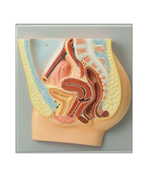
مجسم الجهاز التناسلي الأنثوي على لوحة ثنائي الأبعاد هندي
JOD 15.00 Add to cart المجسمات التشريحيةمجسم الجهاز التناسلي الأنثوي على لوحة ثنائي الأبعاد هندي
-

كاميرا للمايكروسكوب
JOD 265.00 Add to cart المجاهرMVV3000 is a special design USB digital camera, which to be used to convert optical microscope into digital one by adding digital capability. When this digital gadget is added to eyepiece tube or C-mount, you can easily view image on PC screen and to save, and no longer squinting with one eye, just be enjoyable and to share with others.
-
Camera for Microscope 3 Megapixel Model MVV3000
JOD 265.00 Add to cart MicroscopesMVV3000 is a special design USB digital camera, which to be used to convert optical microscope into digital one by adding digital capability. When this digital gadget is added to eyepiece tube or C-mount, you can easily view image on PC screen and to save, and no longer squinting with one eye, just be enjoyable and to share with others.
3 megapixel resolution with free software
-
Female Human Reproductive System 2D (IN)
JOD 15.00 Add to cart Anatomical SeriesFemale Human Reproductive System 2D (IN)
-

Male Human Reproductive System 2D (IN)
JOD 15.00 Add to cart Anatomical SeriesMale Human Reproductive System 2D (IN)
-

Skin Section Model (IN)
JOD 15.00 Add to cart Anatomical SeriesThis economically priced skin section model has been enlarged 70 times and shows a section through three layers of the hair covered skin of the head. It also shows hair follicles with sebaceous sweat glands, receptors, nerves and vessels.
-
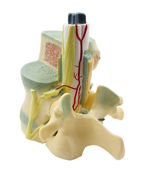
Model Of Spinal Cord And Spine Anatomical
JOD 50.00 Add to cart Educational SeriesStructure – The product consists of adult’s 1st and 2th thoracic vertebrae, 1st and 2th lumbar vertebrae, and partially exposed spinal cord outside the spinal canal. Size: 30cm(Height)
Material – The model is made of polyvinyl chloride (PVC) plastic, which is corrosion resistant, lightweight, and has high strength. So you can use it again and again until you really have the relevant knowledge
Mainly Shows – the external morphology of the 1st and 2th thoracic vertebrae, the 1st and 2nd lumbar vertebrae, the cross-section structure of the spinal cord, the spinal nerve sympathetic trunk, and the vein. So you can to teaching demonstration, which promotes students’ understanding and Improve the quality of teaching
Function – This model is suitable for teaching the morphology of spinal cord and spine in middle schools, colleges and technical colleges. -

مجسم الفكين مع عظام الوجه على قاعدة هندي
JOD 30.00 Add to cart المجسمات التشريحيةShowing various positions of teeth in the upper and lower jaws. Internal wall is removed to show the incisors, permanent canine and permanent premolars, molars, permanent incisors in full view and also the arteries, veins and nerves serving them, on base with key card. Visit www.jaincolab.com for further details and other products related to Physics, Chemistry & Biology Lab.
All Products
Shopping Cart